Raman microscopy and Raman imaging: new tools to explore the Bio-world
Seminario
Pisa, 22 Marzo 2018 9h30 – 11h00 aula seminari Istituto di Biofisica CNR Via Moruzzi 1, 56123, Pisa
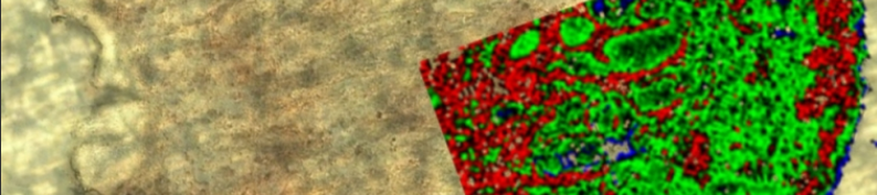
Raman microscopy and related techniques -2D and 3D Raman imaging, TERS (Tip Enhanced Raman Scattering) and SERS (Surface-enhanced Raman spectroscopy) are gaining more and more importance in different research fields amongst which the bio-medical applications, being Raman a highly specific, fast and label free characterization tool.
This seminar aims to propose the last Renishaw technological innovations, with specific focus on the following application areas:
- Tissue imaging
- Cancer diagnosis
- Assessment of pathological state
- Cell type differentiation (stem cells / non-stem cells)
- Live cell studies
- Bacterial species identification
- Cell – drug interaction (uptake pathway, distribution etc)
- In vivo Raman
- Protein structural investigation
- Chemical information of model organism (e.g. nematode worms)
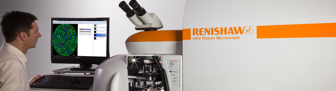
Renishaw is a recognised leader in Raman spectroscopy, producing high performance Raman systems for a range of applications. Renishaw has spearheaded this expansion with innovations such as the award-winning inVia confocal Raman microscope, combined Raman / scanning probe microscope systems, and combined SEM-Raman system. (http://www.renishaw.com/en/raman-spectroscopy–6150)
For enquiries, please contact Mario D’Acunto (mario.dacunto@pi.ibf.cnr.it)